Description
Specification
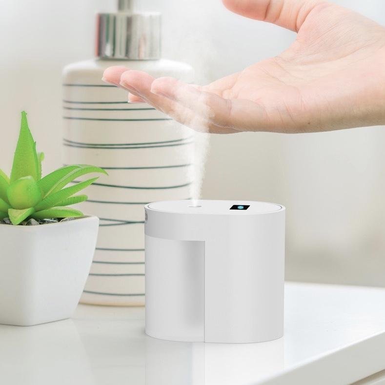
- Size (mm) : 84 * 60 * 76
- Weight: 138 g
- Including spray sterilizer *1, Type C charging cable*1, bilingual manuals (both Chinese and English)*1, cotton swab *2
- The volume of the tank: 100ml
- 15 secs spraying time
Refund and Return Policy
This product is a non-refundable product, please read our return/refund policy for more details.